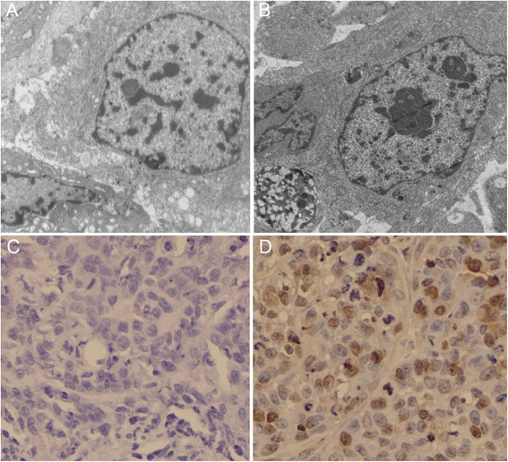
Fig. 7.

Apoptosis array in LSCC xenografts. a Cells in control Hep-2 xenografts had normal membrane, organelles, and nuclear morphology. b Apoptotic morphologies were found in NEAT1 siRNA treated Hep-2 xenografts. c TUNEL staining showed no obvious apoptotic cells in tumors of the control group. d Strong brown staining indicated many TUNEL positive apoptotic cells in NEAT1 siRNA treated Hep-2 xenografts
