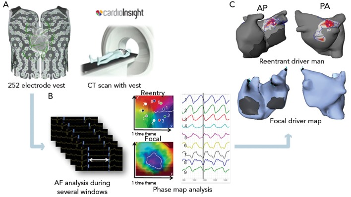
Figure 1: Processing of Body Surface Mapping.

(A) After acquiring CT scan with the 252 electrode vest; (B) multiple atrial fibrillation (AF) windows with subtraction of QRST are analysed to identify consistent drivers by using phase map analysis. (C) The cumulative epicardial driver map is composed on the reconstructed biatrial shell from CT. Density of the driver map is based on the prevalence and trajectory of the reentrant driver core. AP = anterior–posterior ; PA = posterior–anterior
