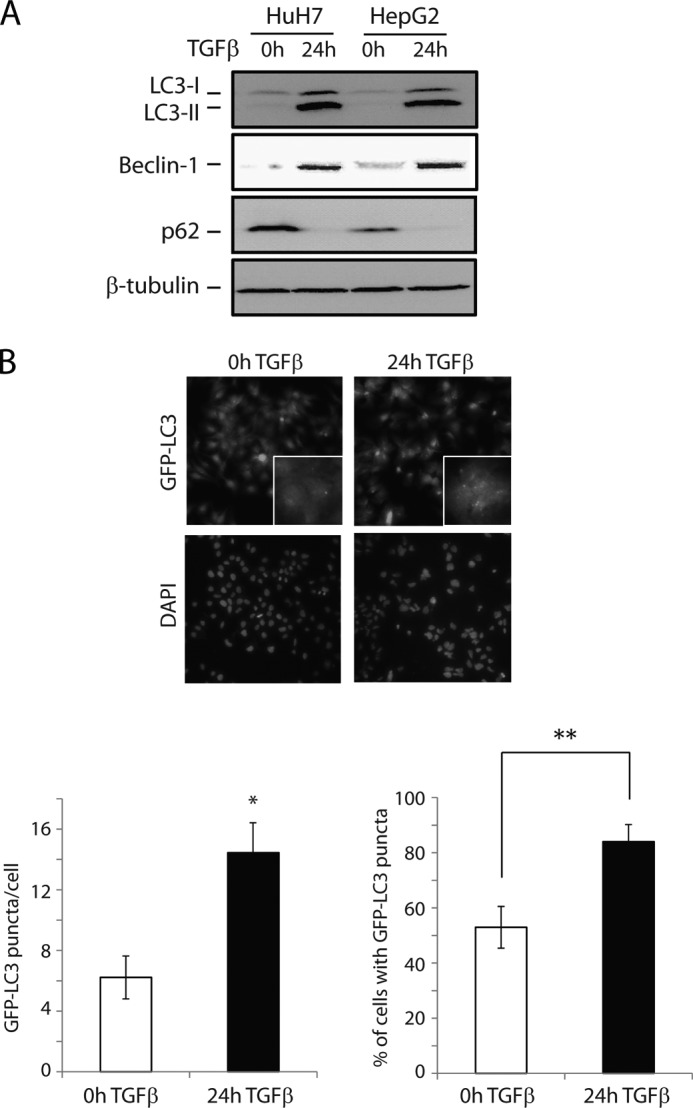
FIGURE 1.

TGFβ induces autophagy in human hepatocarcinoma cell lines. A, immunoblot analysis of the conversion of endogenous LC3 (LC3-I to the more rapidly migrating LC3-II), as well as expression levels of Beclin-1 and p62, in HuH7 and HepG2 cells treated with TGFβ for 24 h. B, HuH7 cells stably expressing GFP-LC3 were treated with TGFβ as indicated, and the relocalization of GFP-LC3 to autophagosomes was detected as punctate formation, visualized by fluorescence microscopy (representative images, left panel). The number of GFP-LC3 puncta per cell and percentage of cells exhibiting more than five GFP-LC3 puncta were quantified (right panel). The data are presented as means ± S.D. **, p < 0.01.
