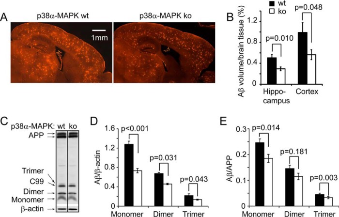
FIGURE 2.
Deletion of neuronal p38α MAPK reduces cerebral Aβ load in APP-transgenic mice. Four-month-old APPtgp38fl/wtNex-Cre+/− (p38α heterozygote, ko) and APPtgp38fl/wtNex-Cre−/− (p38α wild-type, wt) littermate mice were analyzed for cerebral Aβ load after immunofluorescent staining with human Aβ-specific antibody (A). The Aβ volume adjusted by relevant brain volume as estimated with Cavalieri method is reduced in p38α MAPK-deficient APP mice (B; t test, n = 10 and 8 for p38 ko and wt groups, respectively). The cerebral Aβ in these APP-transgenic mice was also evaluated by detecting Aβ in the brain homogenate with quantitative Western blot (C–E, t test, n = 7 and 8 for p38 ko and wt groups, respectively). C, the figure is grouping images from different parts of the same gel.

