FIGURE 3.
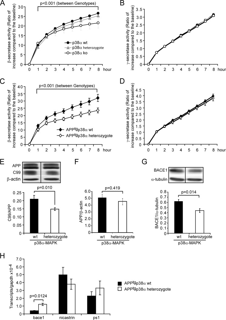
Deletion of neuronal p38α MAPK reduces both β-secretase activity and protein amount in the mouse brain. Four-month-old APP-transgenic (APPtg, C–H) or non-transgenic (A and B), p38fl/flNex-Cre+/− (ko), p38fl/wtNex-Cre+/− (heterozygote), and p38fl/wt or p38fl/flNex-Cre−/− (wt) littermate mice were used to prepare membrane components, brain homogenates, and total RNA. β- and γ-secretase activity was measured by incubating membrane components with fluorogenic β- and γ-secretase substrates, respectively (A–D, two-way ANOVA followed by Bonferroni post hoc test between mouse groups with different genotypes; A and B, n ≥ 4 per group; C and D, n ≥ 10 per group). The protein levels of APP, APP-C99, and BACE1 in the APPtg mouse brain homogenate were determined by quantitative Western blots (E–G, n ≥ 4 per group). Moreover, the transcripts of bace1, nicastrin, and ps1 genes in the APPtg mouse brain were measured with real-time PCR, which showed that transcription of bace1 was up-regulated after deletion of p38α MAPK specifically in neurons (H, t test; n ≥ 4 per group). E or G, the figure is grouping images from different parts of the same gel.

