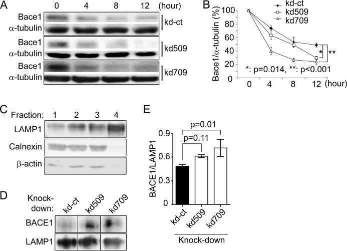
FIGURE 5.
p38α MAPK deficiency promotes lysosomal degradation of BACE1 in SH-SY5Y cells. p38α MAPK knocked down (kd509 or kd709) and control (kd-ct) cells were treated with cycloheximide. Cell lysates were collected 0, 4, 8, and 12 h thereafter. Quantitative Western blot was used to determine the protein levels of BACE1 and α-tubulin (A and B, two-way ANOVA followed by Tukey post hoc test; n = 3 per group). Lysosomes were isolated from different cell lines using Percoll gradient centrifugation. From the top to bottom, 4 times fractions were collected for Western blot detection of LAMP-1, calnexin, and β-actin (C). In the following experiments, the BACE1 protein was detected and quantified in fraction 4 with LAMP-1 as an internal protein-loading control (D and E, one-way ANOVA followed by Tukey post-hoc test; n = 3 per group). A, C, or D, grouping images from different parts of the same gel.

