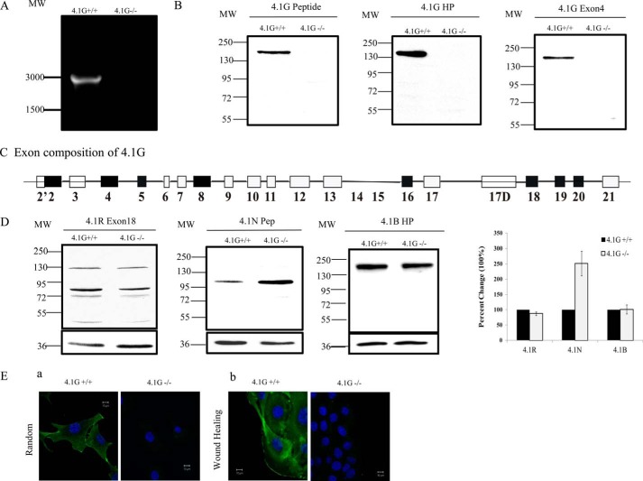
FIGURE 1.
Expression and localization of 4.1G in MEF cells. A, RT-PCR analysis of the expression of 4.1G in 4.1G+/+ and 4.1G −/− MEF cells. MW, molecular weight. B, immunoblot analysis of the expression of 4.1G in 4.1G+/+ and 4.1G−/− MEF cells. Total lysates (35 μg of protein) were probed with polyclonal rabbit antibodies against the 4.1G peptide, the 4.1G head piece, and 4.1G exon4. C, schematic of the 4.1G protein structure and exon organization in MEF cells. D, immunoblot analysis of protein 4.1 members in 4.1G+/+ and 4.1G−/− MEF cells. Total lysates (35 μg of protein) were probed with polyclonal goat antibody against 4.1R exon13 and rabbit antibodies against the 4.1N peptide and 4.1B head piece. Quantitative analysis of immunoblot results from three independent experiments is shown in the right panel. GAPDH was used as a loading control. E, immunofluorescence staining of endogenous 4.1G in randomly migrated and directionally migrated 4.1G+/+ and 4.1G−/− MEF cells. Subconfluent cells were used as randomly migrated cells, and confluent cells were checked 4 h after wounding as directionally migrated cells. Cells were fixed and stained using anti-4.1G-U3 antibody (green) and DAPI (blue).

