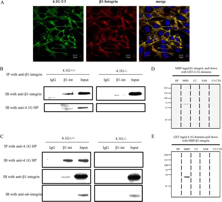
FIGURE 5.
Direct association of β1 integrin with 4.1G. A, immunofluorescence staining showing the co-localization of 4.1G with β1 integrin in confluent 4.1G+/+MEF cells. Cells were fixed and stained using anti-4.1G-U3 antibody (green), anti-β1 integrin antibody (MB1.2), and DAPI (blue). B and C, 4.1G and β1 integrin associate in situ. B, immunoprecipitation (IP) of β1 integrin. β1 integrin (β1 int) was immunoprecipitated from 4.1G+/+ and 4.1G−/− MEF cells using anti-β1 integrin. β1 integrin or 4.1G in the immunoprecipitate was detected using anti-β1 integrin antibody or anti-4.1G HP antibody. IB, immunoblot. C, immunoprecipitation of 4.1G. 4.1G was immunoprecipitated from MEF cells using anti-4.1G HP antibody. 4.1G, β1 integrin, and α6 integrin in the immunoprecipitate were detected using the corresponding antibodies. D, binding of 4.1G to the cytoplasmic domain of β1 integrin. GST-tagged 4.1G-HP, MBD, U2, SAB, and the C-terminal domain (CTD) were incubated for 30 min at room temperature with the MBP-tagged cytoplasmic domain of β1 integrin, and binding was assessed by pulldown assay. β1 integrin binding was detected by blotting with anti-MBP antibody. E, the MBP-tagged cytoplasmic domain of β1 integrin was incubated for 30 min at room temperature with GST-tagged 4.1G HP, MBD, U2, SAB, and C-terminal domains, and binding was assessed by pulldown assay. 4.1G binding was detected by blotting with anti-GST antibody.

