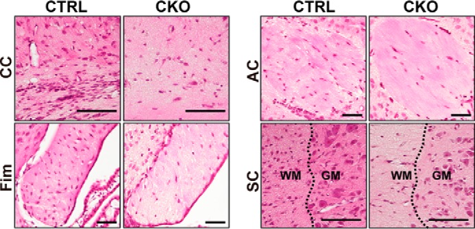
FIGURE 4.

Histological abnormalities of the white matter parts in Prmt1flox/flox; Nes-Cre mice. Hematoxylin and eosin staining images of brains and spinal cords from CTRL and CKO mice at P10 obtained from coronal sections of tissues are shown. CC, corpus callosum; Fim, fimbria; AC, anterior commissure; SC, spinal cord. Dotted lines represent the boundary of white matter (WM) and gray matter (GM). Scale bars represent 100 μm.
