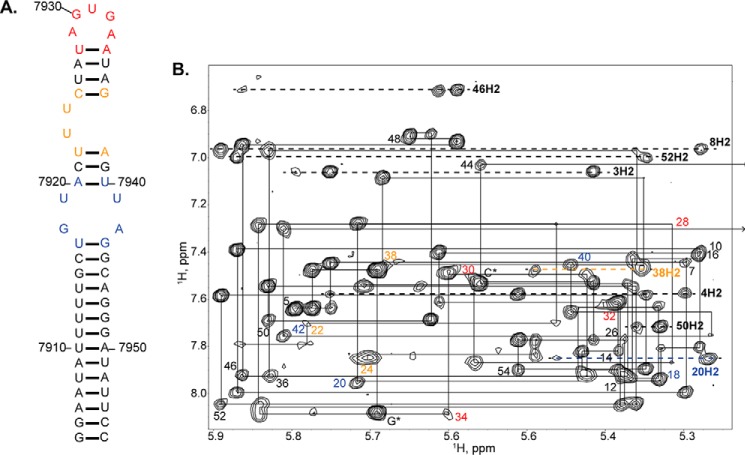
FIGURE 5.
NMR evidence of stable SL1ISS structural elements. A, sequence and secondary structure diagram of SL1ISS where nucleotides are color-coded according to structural elements: black, helical regions; blue, 2X2 internal loop; orange, UU bulge; and red, 5-nt AGUGA apical loop. B, representative 1H-1H NOESY (900 MHz and τm = 250 ms) spectrum collected in 5 mm K2HPO4 (298 K and pH 6.5) and 100% D2O. Assignments of the intranucleotide NOE (H8/H6-H1′) interactions are indicated for every other nucleotide. Numbers are colored according to secondary structural elements shown in A. 7,900 should be added to the assignment labels to obtain the native HIV-1Bru numbering. Note that G* corresponds to the non-native guanosine added to increase transcription yields. Lines denote the sequential walk pathway, and the arrow indicates that the G7943 H1′ chemical shift is upfield (∼5 ppm) relative to all other H1′ chemical shifts.

