
FIGURE 3.
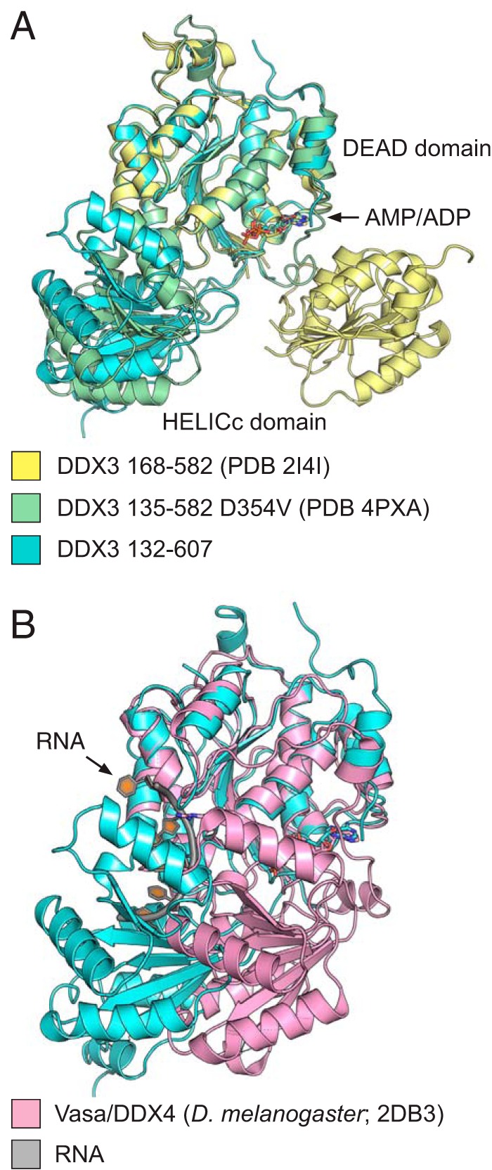
The 2.2 Å crystal structure of the conserved core of wild-type DDX3. A, the structure of DDX3 132–607 bound to AMP (blue) is shown along with the structure of DDX3 135–582 D354V (green; PDB 4PXA) and 168–582 (yellow; PDB 2I4I). Structures are aligned by the DEAD domain, highlighting the rotation of the C-terminal HELICc domain between the three structures. B, the partially closed state of DDX3 (blue) clashes with the RNA-binding site based on a comparison with the DEAD-box protein Vasa bound to RNA (Vasa: pink; RNA: gray).