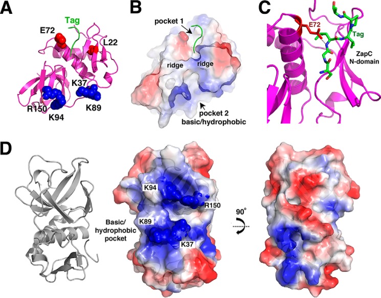
FIGURE 3.
Pockets on the ZapC structure. A, ribbon diagram of ZapC showing relative location of pockets in the N-domain and C-domain. Residues previously identified as important in ZapC function/FtsZ binding, Glu-72 and Leu-22 (colored red), that line the N-domain pocket are shown as spheres. Basic residues that surround the larger C-domain pocket are shown as spheres and are colored blue. Also shown is the N-terminal tag that was found docked into the N-domain pocket (green). B, surface representation of ZapC shown in the same orientation as A and colored according to electrostatic potential, with blue and red representing electropositive and electronegative regions, respectively. This figure underscores the strong electropositive nature of the sides of the C-domain pocket. This figure also underscores the hydrophobic nature of the pocket cavities (indicated as white surfaces). C, close up of the interaction between the tag residues docked in the N-domain pocket and the side chain of Glu-72. D, focus on the C-domain pocket and the location of the basic residues that line the pocket, which were mutated to assess their roles in ZapC function. Left is the ribbon diagram to orient the location of the pocket, and center is the electrostatic surface. Right is the same electrostatic figure but rotated by 90° to underscore that this surface has neither strong charge characteristics nor does it contain a deep pocket.

