FIGURE 5.
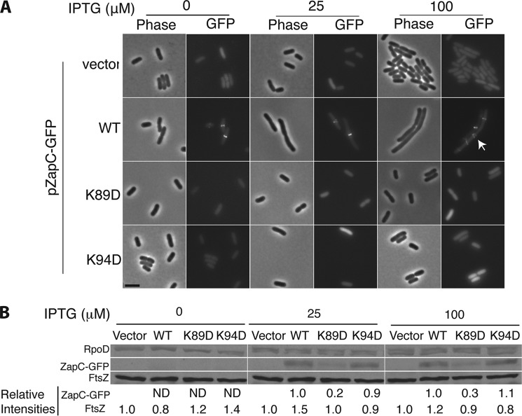
ZapC(K89D) and ZapC(K94D) mutants were unable to localize GFP to midcell. A, representative phase and fluorescence images of WT (MG1655) cells expressing ZapC-GFP (KHH236) or mutant ZapC-GFP fusions (KHH237 and KHH239) were grown and visualized as described in “Materials and Methods.” The arrow in the image panel expressing WT ZapC upon 100 μm IPTG induction indicates aberrant localization of the ZapC-GFP fusion. Bar = 5 μm. B, quantitative immunoblotting of FtsZ, ZapC and mutant ZapC proteins upon overexpression of ZapC- or mutant ZapC-GFP fusions. Whole cell proteins from equal quantities of each strain (KHH236, KHH237, and KHH239) were separated by SDS-PAGE and immunoblotted with anti-GFP, anti-FtsZ, or anti-RpoD antibodies. A representative blot (of five independent trials) with relative intensity values is reported. ND = signal not detectable.

