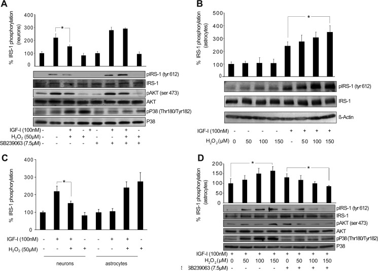
FIGURE 2.
AKT activation by IGF-I in astrocytes is not affected by p38 redox activation. A, neurons treated with IGF-I (100 nm) showed a significant up-regulation of phospho-IRS-1 (pIRS-1) (Tyr-612) and phospho-AKT (pAKT) (Ser-473) levels. Co-administration of H2O2 (50 μm) increased phospho-p38 (pP38) (Thr-180/Tyr-182) levels and prevented IGF-I effects on phospho-IRS-1 and phospho-AKT levels (*, p < 0.05 versus IGF-I-treated cells; n = 3). Neuron pretreatment with the p38 inhibitor SB239063 (7.5 μm) prevented H2O2-mediated p38 activation and restored IGF-I effects on phosho-IRS-1 and phospho-AKT. β-Actin served as a loading control. B, astrocytes treated with IGF-I (100 nm) showed an up-regulation of phospho-IRS-1 (Tyr-612) levels. Co-administration of different doses of H2O2 (50–150 μm) did not prevent this up-regulation. On the contrary, the highest dose of H2O2 (150 μm) significantly enhanced IGF-I effects (*, p < 0.05 versus IGF-I-treated cells; n = 4). β-Actin served as a loading control. C, both neurons and astrocytes treated with IGF-I (100 nm) showed an up-regulation of phospho-IRS-1 (Tyr-612) levels. However, although in neurons co-administration of H2O2 (50 μm) prevented IGF-I effects on phospho-IRS-1, in astrocytes this effect was not present (*, p < 0.05 versus IGF-I-treated neurons; n = 3). D, H2O2 (150 μm) administration significantly enhanced phospho-IRS-1 (Tyr-612) up-regulation induced by IGF-I (100 nm) in astrocytes (*, p < 0.05 versus IGF-I-treated cells; n = 4). Astrocyte pretreatment with the p38 inhibitor SB239063 (7.5 μm) prevented H2O2-mediated p38 activation, but it did not affect increased phosho-IRS-1 levels by IGF-I (100 nm) except with H2O2 (150 μm), which significantly reduced them (*, p < 0.05 versus IGF-I-treated cells in the presence of SB239063 inhibitor; n = 3). β-Actin served as a loading control. Error bars represent S.E.

