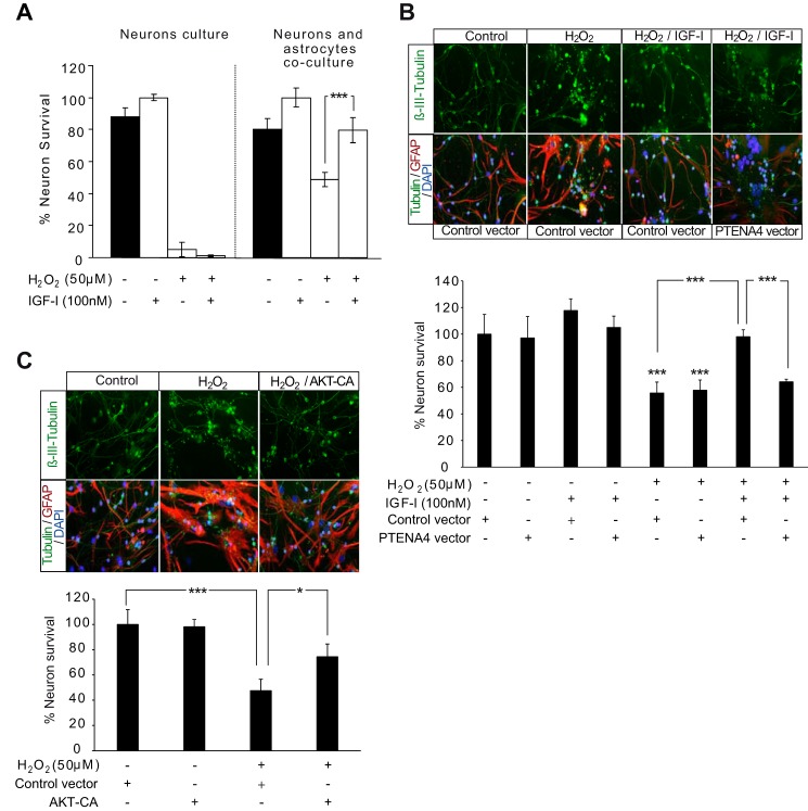
FIGURE 6.
AKT activation by IGF-I is involved in neuroprotection by astrocytes. A, neuron survival was determined in cultures of neurons alone or in combination with astrocytes. These cultures were treated with H2O2 (50 μm), IGF-I (100 nm), or the combination. Quantification of neuron survival was assessed by counting the total number of viable β-III-tubulin+ (neuronal marker) cells determined by plasma membrane integrity and the absence of nuclear alterations detected with DAPI. The percentage of viable neurons was expressed relative to IGF-I-treated cultures. Deleterious effects of H2O2 were much stronger in neuron cultures alone than in combination with astrocytes. IGF-I co-administration increased neuron survival only in co-cultures with astrocytes (***, p < 0.001 versus H2O2-treated co-cultures; n = 3). B, upper, representative photomicrographs of co-cultures of neurons with astrocytes immunostained with β-III-tubulin (neurons), GFAP (astrocytes), and DAPI (cell nuclei). Astrocytes were transfected with PTENA4 mutant or a control construct prior to co-culture with neurons and treatment with H2O2 (100 μm), IGF-I (100 nm), or both. Lower, quantification of neuron survival was assessed by counting the total number of viable β-III-tubulin+ (neuronal marker) cells determined by plasma membrane integrity and the absence of nuclear alterations detected with DAPI. The percentage of viable neurons was expressed relative to vehicle treatments. H2O2 exposure significantly reduced neuron survival both in co-cultures with PTENA4 astrocytes and with control astrocytes (***, p < 0.001 versus control co-cultures; n = 3). IGF-I co-administration significantly prevented H2O2 effects on neuronal survival in co-cultures with control astrocytes (***, p < 0.001 versus H2O2-treated co-cultures with control construct astrocytes; n = 3), whereas in co-cultures with PTENA4-transfected astrocytes, IGF-I effects were abolished (***, p < 0.001 versus H2O2 + IGF-I-treated co-cultures with control construct astrocytes; n = 3). C, upper, representative photomicrographs of co-cultures of neurons with astrocytes immunostained with β-III-tubulin, GFAP, and DAPI. Astrocytes were transfected with an AKT-CA mutant or a control construct prior to co-culture with neurons and treatment with H2O2 (100 μm). Lower, H2O2 exposure significantly reduced neuron survival in co-cultures with control astrocytes (***, p < 0.001 versus control co-cultures), whereas in co-cultures with AKT-CA-astrocytes, the H2O2 effect was partially prevented (*, p < 0.05 versus H2O2-treated co-cultures with control construct astrocytes). Error bars represent S.E.

