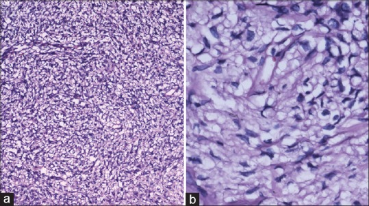
Figure 3.

(a) Tumor revealing patternless fibroblastic proliferation and vascular channels with pericytomatous arrangement (H and E, ×100) (b) spindle cells growing in a haphazard manner in a variable cellular stroma described as “patternless pattern” (H and E, ×400)
