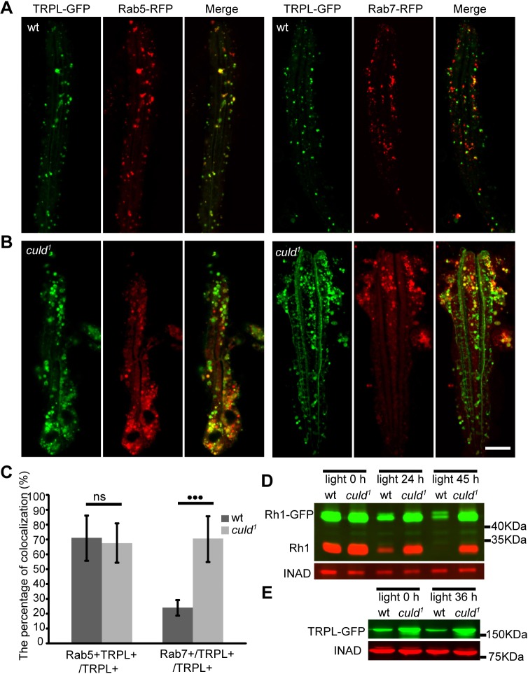
Fig. 6.
Defective light-dependent turnover of Rh1 and TRPL in culd1 flies. (A,B) Colocalization between (A) Rab5–RFP (red) and (B) Rab7–RFP with TRPL–GFP (green) in ommatidia. Three-day-old flies with ninaE-trpl-gfp and ninaE-rab5-rfp or ninaE-rab7-rfp transgenes in either wild-type or culd1 background were placed in the dark for 12 h, before exposed to orange light for 2 h. Scale bar: 10 µm. (C) Quantification of the percentage of TRPL and Rab5 double-positive vesicles among TRPL-positive vesicles (TRPL+Rab5+/TRPL+) and the percentage of TRPL and Rab7 double-positive vesicles among TRPL-positive vesicles (TRPL+Rab7+/TRPL+) are shown for the experiments in A and B. At least nine ommatidia were quantified for each sample as described in Materials and Methods. Error bars indicate s.d. ***P<0.001; ns, not significant (unpaired t-test). (D) Light-induced Rh1 degradation was blocked in culd1 flies. Western blots of heads were from ninaE-rh1-gfp; cn bw; culd1/TM6B (wt) and ninaE-rh1-gfp; cn bw; culd1 (culd1) flies exposed to blue light for the indicated periods of time. INAD served as a loading control, and protein extracts from half of a fly head were loaded for each line. (E) Western blot analysis of heads from ninaE-trpl-gfp (wt) or cn bw; ninaE-trpl-gfp, culd1 (culd1) flies that were exposed to orange light for indicated periods of time. Protein extracts of one head were loaded for each line. Two-day-old wt or culd1 flies with white eyes were used.

