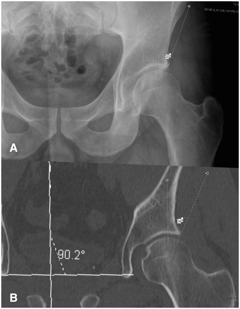
Fig. 2.
Representative example of lateral acetabular dysplasia. The measured LCEA is (A) 20° on AP pelvic radiography, compared with (B) 24° on coronal CT. These measurements confer a clinical diagnosis of frank hip dysplasia and borderline hip dysplasia, respectively—a discrepancy that could alter the course of operative treatment.

