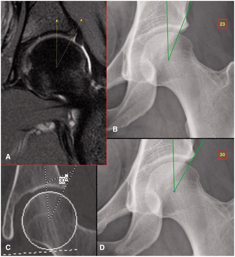
Fig. 3.
Representative example in borderline dysplastic hip. (A) Coronal MRI of the left hip shows the attachment point of the labrum to the lateral margin of the acetabular roof. (B) Measurement of the LCEA (of Ogata) yields a value of 23° when using a terminal endpoint at the weight-bearing area of the acetabulum that, as seen by the MRI, is located at the medial base of the labrum. (C) Measurement of the LCEA (of Wiberg) on coronal CT involves a terminal endpoint located at the far lateral acetabular rim, yielding a value of 30°. (D) Measurement of the LCEA on plain radiography using a technique analogous to that performed on coronal CT. The difference between LCEA measurements in panels B and D is attributable to a bony area which functions as the labral base but does not come into contact with the femoral head and thus does not contribute directly to the acetabular coverage.

