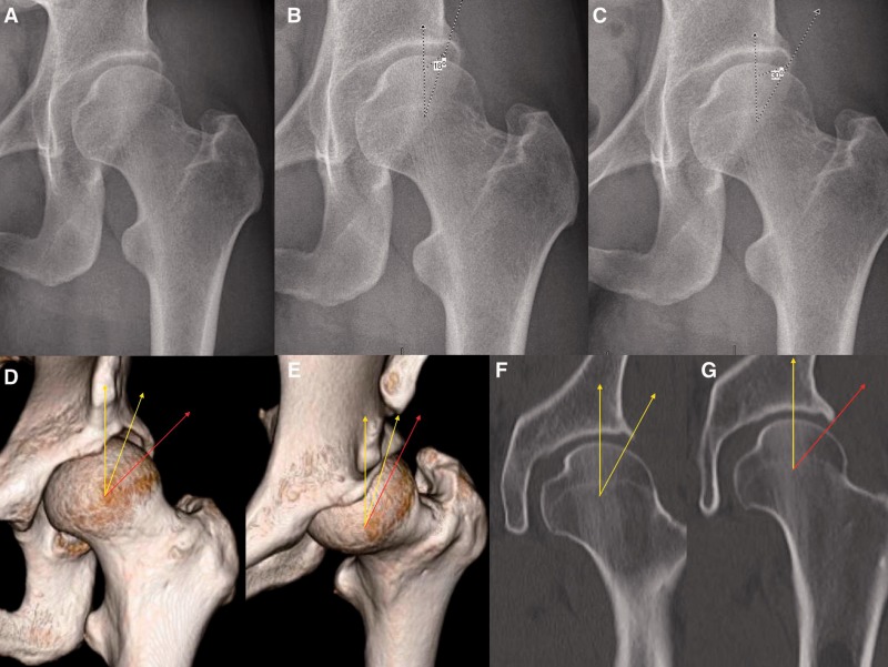
Fig. 4.
Potential challenges associated with measurement of LCEA in dysplastic hips. (A) AP pelvic radiograph of the left hip; (B) measurement of the LCEA with a terminal endpoint at the intersection of acetabular rim and anterior acetabular wall yields a value of 18°, indicating acetabular dysplasia, (C) while measurement with a terminal endpoint at (what seems to be) the lateral rim yields a value of 30°; (D, E) 3D CT enables superior visualization of bony morphology. Note that the lateral border of the anterior inferior iliac spine landmark should be positioned at 1 o’clock to ensure measurement of the LCEA to the true anterolateral rim (yellow) at the 12 o’clock position. Failure to do so can potentially result in erroneous measurement of the LCEA with an endpoint on the posterior acetabular wall that does not contribute to anterolateral coverage (red). Corresponding measurement on coronal CT views are shown in panels (F) and (G).

