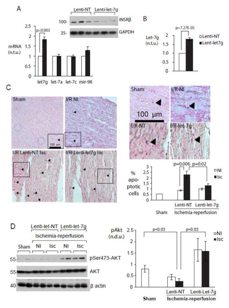
Figure 5. miR-let-7g protects myocardium from acute ischemia-reperfusion injury by increasing AKT activation.
A. Lentiviral transduction of miR-let-7g in myocardium in vivo. Transcripts levels of miR-let-7 family members and protein levels of INSRβ, a validated miR-let-7 target, were assayed in whole heart tissue using RT-PCR and immunoblot 10 days after lentiviral vector injection. (Left) Transcript levels relative to lenti-NT-transduced animals. (Above) Representative Western blot. B. miR-let-7g expression is sustained to adulthood. MiR-let-7g transcript levels were measured in whole hearts 9–10 weeks after transduction (n=3-5). C. Increasing miR-let-7g reduces apoptosis in ischemia-reperfusion injury. Sections from sham-operated hearts, and from non-ischemic and ischemic zones of NT- and miR-let-7g-transduced mice were examined for apoptotic cells following 60 minutes of ischemia and 24 hours of reperfusion as described in Experimental Procedures. (Left) Representative sections and (top) enlargements. Sham = sham-operated. I/R NI = Ischemia-reperfusion, non-ischemic zone. I/R-NT, I/R-let-7g = I/R + non-targeting and let-7g vectors respectively. (Bottom right) Quantitation of apoptotic nuclei (n=2500–4000 nuclei) in n = 4 (NT) or 5 (let-7g) individual mice. D. AKT is activated in vivo with miR-let-7g overexpression. Non-ischemic and ischemic zones of hearts from NT and miR-let-7g-transduced animals were assayed for pSer473-Akt, total Akt and β-actin. (Left) Representative Western blots. (Right) Graph summarizes data from n=4 (NT) and n=5 (let-7g) mice.

