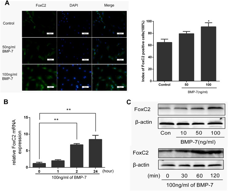
Fig 4. Immunohistochemical staining of NP cells against FoxC2.
(A) After 1-day serum deprivation, NP cells were treated 50 ng/ml or 100 ng/ml BMP-7 in serum-free medium for 2h. NP cells were Immunohistochemical stained against FoxC2. Nuclei were stained with DAPI, shown in blue. Images were acquired using laser scanning confocal microscopy under a 40×objective. FoxC2-positive percentages in cultured NP cells 2h after treated with 50 ng/ml or 100 ng/ml BMP-7 or control was under analysis. (B) NP cells were stimulated by BMP-7 (100 ng/ml) for the indicated periods of time. And then expression level of FoxC2 was measured by real-time RT-PCR. (C) NP cells were stimulated by 10, 50, and 100 ng/ml BMP-7 for 2h or NP cells were stimulated by BMP-7 (100 ng/ml) for the indicated periods of time. Cell lysates were then prepared and analyzed by western blotting with specific anti-FoxC2 antibody. β-actin was used as a control for normalization. Error bars represent SD. As compared with control, * indicates p<0.05, ** indicates p<0.01.

