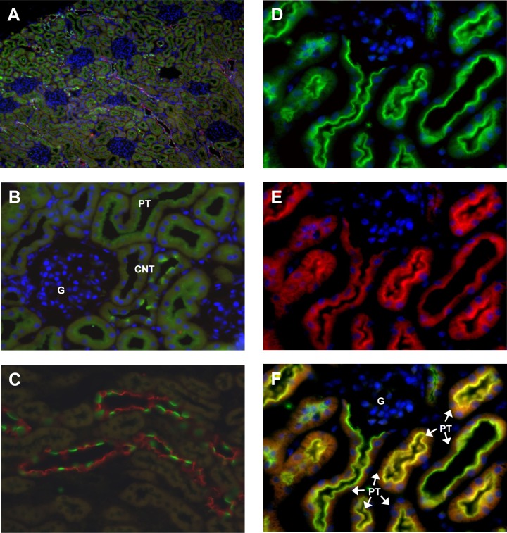
Fig 2. Localization of the (P)RR/Atp6ap2 in mouse kidney.
Immunoflourescence staining for the (P)RR/Atp6p2 in mouse kidney (green). (A) Overview showing (P)RR/Atp6ap2 (green), the principal cell specific AQP2 water channel (red), and nuclei (blue), original magnification 40 x. (B) A cortical field with glomerulus (G), and (P)RR/Atp6ap2 staining (green) in the proximal tubule (PT) and connecting tubules (CNT), 400 x magnification. (C) Cortical and outer medullary collecting duct stained for (P)RR/Atp6ap2 (green) and the principal cell specific AQP2 water channel (red), 400 x magnification. (E,D,F) (P)RR staining (D: green) was detected in the proximal tubule (PT) in the brush border membrane and colocalized with the a4 H+-ATPase subunit (ATP6V0A4)(E: red) as indicated by the yellow color (F), original magnification 400x.

