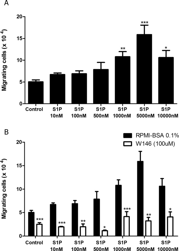
Fig 4. High S1P concentrations induce S1P1-dependent fugetaxis of CEM cells.
(A) CEM cells were serum-starved for 2 h, applied to Transwell™ chambers containing different S1P concentrations and incubated for 4 hours. S1P was added to the upper chamber to evaluate fugetaxis; only RPMI-BSA 0.1% was added to the bottom chambers. Results were analyzed by One-way ANOVA, followed by Tukey post-test (n = 3). (B) CEM cells were serum-starved for 2 h and pre-treated or not with W146 (100 μM). Cells were then applied to Transwell™ chambers containing different S1P concentrations and incubated for 4 hours. S1P was added to the upper chamber to evaluate repulsive responses. Black bars correspond to T-ALL blasts pre-treated with RPMI-BSA 0.1% alone and white bars correspond to T-ALL blasts pre-treated with W146. Results were analyzed by unpaired Student’s t test. Results are expressed as mean ± SEM and differences were considered statistically significant when * p˂0.05, ** p ˂0.01 or *** p ˂0.001 (n = 3).

