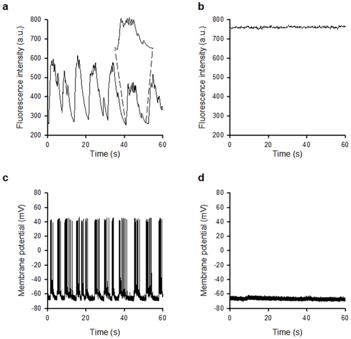
Fig 6. Widefield two-photon excitation for recording synaptic activity.

(a) Fluorescence recording at a frame rate of 10 Hz shows spontaneous changes in fluorescence intensity in live neuronal cell bodies loaded with Fluo-4 AM, which were abolished in the presence of the glutamatergic antagonists DL-AP5 and NBQX (b). The elevated Ca2+ level seen in Fig 5b fits with the observation of an overall increase in two-photon excited fluorescence signal over time at an image acquisition rate of 100 Hz shown in Fig 4. The expanded region of (a) shows the envelope of events indicating synaptically-driven activity. (c) Whole-cell current clamp recordings from hippocampal neurones revealing spontaneous action potential firing which is abolished in the presence of DL-AP5 and NBQX (d).
