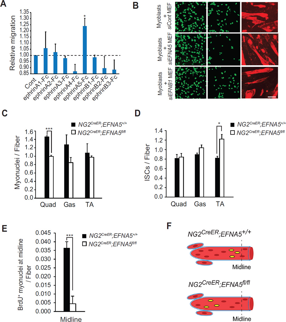
Figure 4. EFNA5 from NG2+ Cells Mediates Myoblast Migration in vitro and in vivo.
(A) A Boyden chamber was used to measure migration of myoblasts when exposed for 24h to different recombinant Fc-clustered ephrins. (B) A two-chamber assay system was used to measure migration of C2C12-H2B:GFP myoblasts toward myotubes when co-cultured with MEFs transfected with siRNAs against EFNA5 or EFNB1. Relative migration is shown as mean ± s.e.m. Myonuclei number per fiber (C) and DAPI+ ISCs (D) was measured from Quad, Gas and TA muscles from P30 NG2CreER;EFNA5+/+ and NG2CreER;EFNA5fl/fl mice. (E) BrdU was injected in NG2CreER;EFNA5+/+ and NG2CreER;EFNA5fl/fl mice and at P30 muscle sections were analyzed for the sublaminar localization of BrdU+ cells at the midline of EDL muscles. (F) A model for BrdU+ cell accretion in muscles from NG2CreER;EFNA5+/+ and NG2CreER;EFNA5fl/fl mice. Scale bars = 100 µm for (B) *p < 0.05, ***p < 0.001 by the student’s t-test. See accompanying Figure S4. Error bars, SEM

