
Fig. 4.
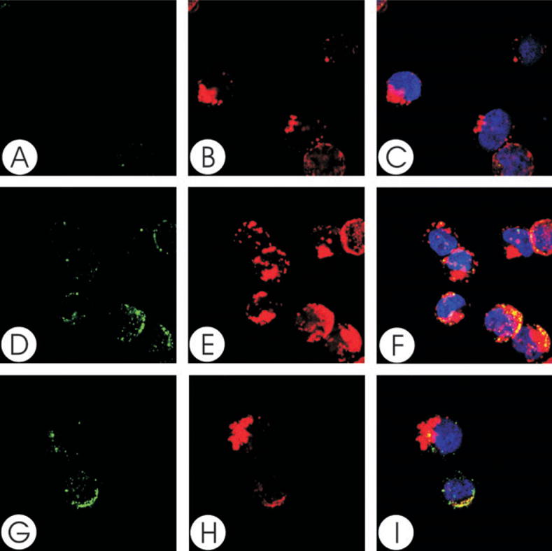
Colocalization of CdtC with GM1. Jurkat cells were treated with CTB conjugated to AlexaFluor 647 and patched with anti-CTB sera. The cells were then treated with Cdt holotoxin and stained with control IgG (A–C) or anti-CdtC (mAb112; D–I) as described in Experimental procedures and assessed using laser confocal microscopy. Images of FITC fluorescence (A, D and G) and AlexaFluor 647 fluorescence (B, E and H) are shown as well as merged images (C, F and I) showing both FITC (green) and AlexaFluor 647 (red) fluorescence along with DAPI-stained nuclei; colocalization is shown in yellow. Analysis of the images indicates that >88% of CdtC fluorescence colocalizes with CTB fluorescence. Results are representative of three experiments.