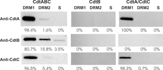
Fig. 6.

Western blot analysis of Cdt peptides associated with DRMs. Jurkat cells were treated with Cdt holotoxin, CdtB peptide or both CdtA and CdtC for 2 h; DRMs were isolated as described in Experimental procedures. DRM1, DRM2 and the soluble fraction were analysed for the presence of each Cdt peptide by Western blot analysis using mAb specific for CdtA, CdtB and CdtC. The immunoblots were analysed by digitized scanning densitometry; the numbers represent the relative distribution (%) of each subunit within DRM1, DRM2 and the soluble (s) fractions respectively. Results are representative of three experiments.
