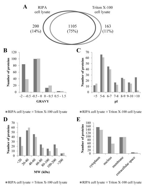
Figure 5.
Comparison of the number, biophysical characteristics and expected cellular location of MDDC proteins identified from RIPA cell lysate and Triton X-100 cell lysate. Both cell lysates were precipitated by acetone and the acetone-washed pellets were solubilised by 1% Na-DOC in 0.5M TEAB dissolution buffer (workflow pAwA-sTD). (A) Distribution of all proteins after merging four experimental replicates for each workflow; Uniquely identified proteins in each sample type distributed according to their (B) GRAVY index, (C) calculated isoelectric point (pI), (D) average molecular weight (MW) and (E) expected subcellular localisation based on associated cellular component GO terms.

