
Fig. 1.
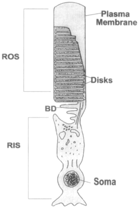
Schematic representation of rod photoreceptor cell. Rod cells are divided into two functionally distinct regions: the rod inner segment (RIS) containing the nucleus and intracellular organelles, and the rod outer segment (ROS) containing disks surrounding by the plasma membrane. BD, basal disks. (See also color insert.)