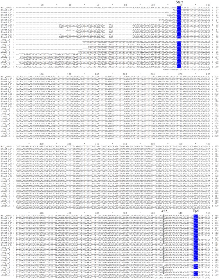
Fig 9. Alignment of HbI cDNA sequences obtained by RT-PCR.
HbI cDNA was amplified with forwards primers designed from short and long variant and a common reverse primer, the sequences are named short and long, respectively. Samples from 1 to 3 correspond to RNA isolated from clams kept in a fish tank and samples 4 to 6 correspond to RNA isolated from clams harvested from their natural environment. F indicates that the sequence was obtained with the corresponding forward primer used for the cDNA amplification and R indicates that the sequence was obtained with the reverse primer. The first three bases coding for the first amino acid at the protein level are marked as start and the last marked as end, both highlighted in blue. Each position that presented different nucleotides are indicated by the number of this position in the reported mRNA for each Hb. The alignments were generated using Clustal W [23] and visualized and formatted with GeneDoc [35].

