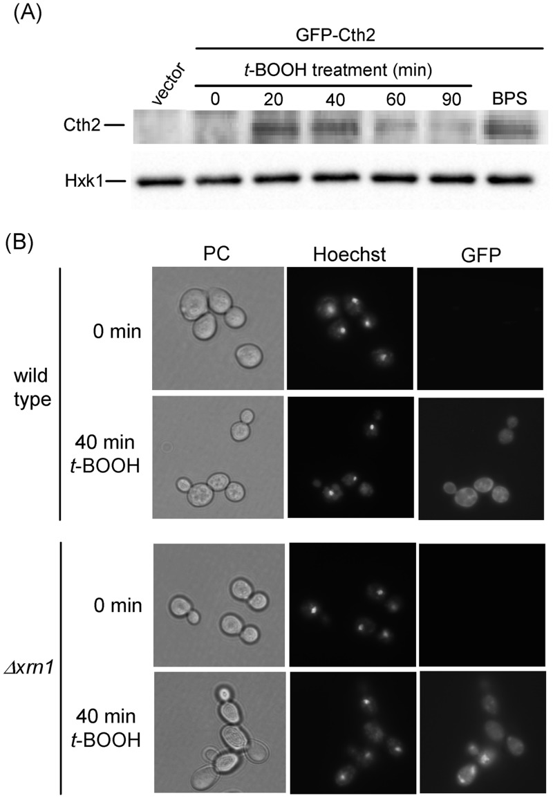
Fig 3. Expression of the Cth2 protein upon oxidative stress.
(A) Western blot analysis of GFP-Cth2 levels in MML1081 (Δcth1 Δcth2) cells transformed with plasmid pRS416-GFP-CTH2 or with vector pRS416, growing exponentially in SC medium at 30°C. Cultures were treated with 0.4 mM t-BOOH for the indicated times or with 100 μM BPS for 6 hours. Anti-GFP antibody was employed for western blot of protein extracts (upper panel), and anti-hexokinase 1 (Hxk1) was employed as loading control (lower panel). (B) Fluorescence microscopy of W303-1A (wild type) or MML1186 (Δxrn1) cells transformed with pRS416-GFP-CTH2, growing exponentially in SC medium at 30°C and then treated for the indicated times with 0.4 mM t-BOOH. Prior to GFP fluorescence analysis samples were stained with Hoechst for nuclei localization. The corresponding phase contrast fields (PC) are also shown.

