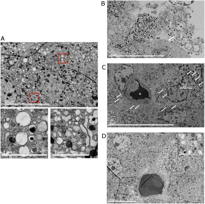
Fig 2. Nanotomy of collagenase-isolated pancreatic rat islets.
(A) A cell with severe stress. The red-boxed areas show swollen vesicles and dilated stressed ER. These vesicles can be desintegrated mitochondria or empty insulin granula indicating a lack of insulin production. (B) During the isolation process the enzymes can disintegrated the cell-membrane causing the organelles and insuline granules to float around in remnants of cytosol, not being contained by a cell membrane. The arrow indicates an intact cell membrane. (C) An islet isolated with batch 1 collagenase. The structure with A depicts a capillary. The β-cells surrounding the capillary are well granulated with insulin granula (the arrows indicate a few classical examples, see insert). The β-cells are polarized. The majority of granula can be found on the site of the β-cell adjacent to the blood vessels. For full resolution, visit http://www.nanotomy.org/deVos/2013-319a/1.html. (D) An islet isolated with collagenase batch 2. The β-cells contain much less insulin granules (see insert) than the batch 1 β-cells and are not polarized. Note that the few granula are found scattered throughout the cell. For full resolution, visit http://www.nanotomy.org/deVos/2013-320a/1.html.

