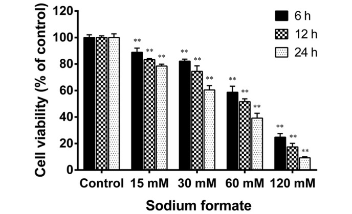
Figure 1.

Sodium formate reduced the viability of 661W cells. The 661W cells were treated with 0, 15, 30, 60 or 120 mM sodium formate. Following treatment for 6–24 h, 20 µl (3-(4,5-Dimethylthiazol-2-yl)-2,5-Diphenyltetrazol ium Bromide was added to each well, followed by an incubation for an additional 4 h. The data revealed the relative proportion of viable cells (%) in the sodium formate-treated and control groups. The results are expressed as the mean ± standard deviation of three independent experiments (*P<0.05; **P<0.01, sodium formate-treated vs. control).
