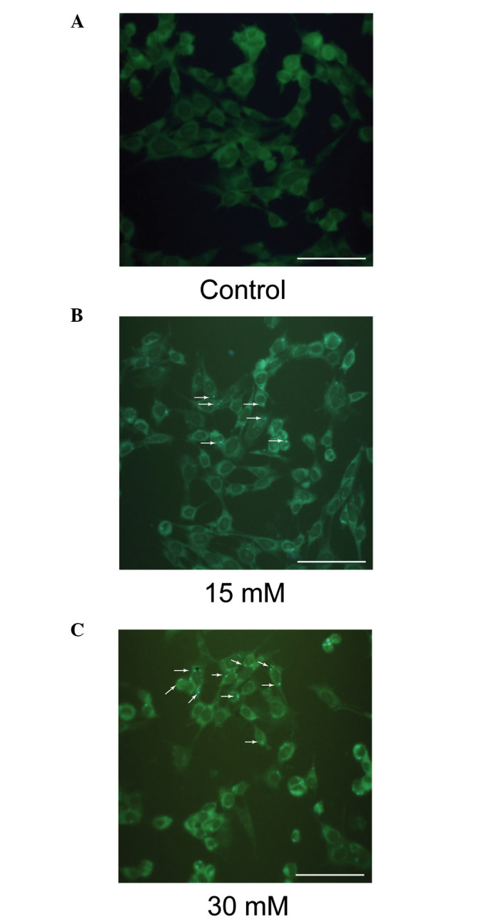
Figure 8.

Sodium formate exposure induces the formation of MDC-positive autophagosomes in 661W cells. The 661W cells were exposed to (A) 0 (control), (B) 15 or (C) 30 mM sodium formate. Autophagic vacuoles were labeled with 0.05 mM MDC in phosphate-buffered saline at 37°C for 10 min. The fluorescence density and the MDC-labeled particles in the 661W cells were greater in the group subjected to sodium formate treatment for 6 h compared with the control group. MDC-labeled vesicles are indicated by arrows (Scale bar=50 µm). MDC, monodansylcadaverine.
