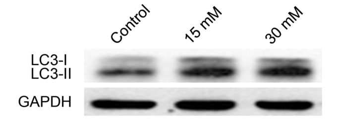
Figure 9.

Cell lysates were analyzed by western blotting with anti-LC3 and GAPDH antibodies. GAPDH was used as an internal control to normalize the quantity of proteins applied in each lane. The protein expression of LC3-II increased in the 661W cells treated with 15 or 30 mM sodium formate. GADPH, glyceraldehyde-3-phosphate dehydrogenase; LC3, microtubule-associated protein 1A/1B-light chain 3.
