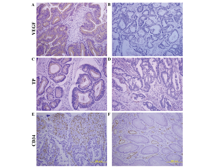
Figure 1.
Immunohistochemical expression patterns of VEGF, TP and CD34 for MVD. (A) High VEGF expression, ≥50% staining in tumor cells. (B) Low VEGF expression, <50% staining in tumor cells. (C) High TP expression, ≥50% staining in tumor cells. (D) Low TP expression, <50% staining in tumor cells. (E) High MVD, ≥81.33/field. (F) Low MVD, <81.33/field. Magnification, ×20. VEGF, vascular endothelial growth factor; TP, thymidine phosphorylase; CD34, cluster of differentiation 34; MVD, microvessel density.

