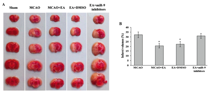
Figure 1.
Effect of EA on infarct volume and morphological structure of rat brains. (A) 2,3,5-Triphenyltetrazolium chloride staining indicating cerebral infarct volumes of Sham, MCAO, MCAO + EA, EA + DMSO and EA + miR-9 inhibitors groups. (B) Bar graph showing the percentage of total brain volume in each group (n=4; *P<0.05, vs. the MCAO and the EA + miR-9 inhibitors groups). MCAO, middle cerebral artery occlusion; EA, electroacupuncture; miR-9, microRNA-9; DMSO, dimethyl sulfoxide.

