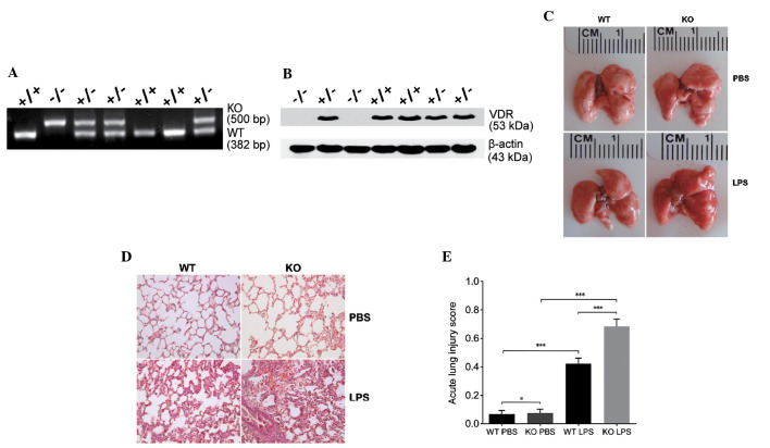
Figure 1.
VDR KO mice exhibit more severe acute lung injury (ALI) than CDR WT mice after LPS treatment. (A) Mouse genotyping of VDR [WT(+), KO(−)]. (B) Western blotting of lung lysates from WT (VDR+/+), heterozygous (VDR+/−) and KO (VDR−/−) mice with anti-VDR antibody. Following exposure to LPS or PBS, the (C) morphology and (D) histology of lung tissue sections from WT and VDR KO mice were investigated. Hematoxylin and eosin stain; magnification, x200. (E) ALI scores according to microscopic examination for the two mouse models. Data are presented as mean ± standard deviation (n=5). #P>0.05, ***P<0.001. KO, knockout; WT, wild-type; VDR, vitamin D receptor; PBS, phosphate buffered saline; LPS, lipopolysaccharides.

