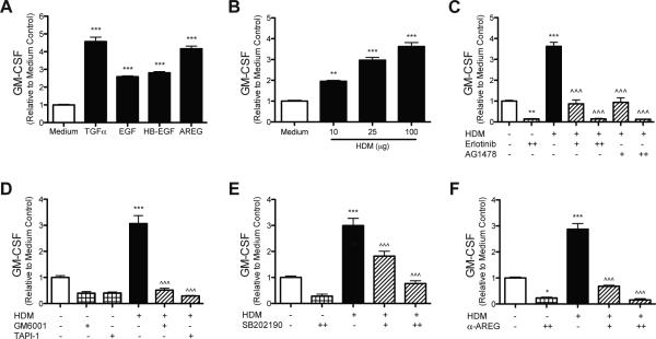
Figure 1. EGFR signaling, protease/TACE activity, and p38 MAPK signaling are required for GMCSF production by HBEC stimulated with HDM.
HBEC were stimulated with (A) equimolar concentrations (72 nM) of TGFα, EGF, HB-EGF, and AREG; (B) HDM (10, 25, or 100 μg dry weight); (C) HDM (100 μg) with or without EGFR inhibitors Erlotinib (0.1 μM [+] or 0.25 μM [++]) or AG1478 (0.25 μM [+] or 0.5 μM [++]); (D) HDM (100 μg) with or without the protease inhibitor GM6001 (20μM) or the TACE inhibitor TAPI-1 (20 μM); (E) HDM (100 μg) with or without the p38 inhibitor SB202190 (2 μM [+] or 5 μM [++]); (F) HDM (100 μg) with or without neutralizing antibodies to AREG (0.3 μg/mL [+] or 1 μg/mL [++]). GMCSF was measured in the media by ELISA 24 hours after stimulation (A-F), and statistical significance was determined by comparing samples to the medium only group (* p<0.05, ** p<0.01, *** p<0.001) or compared to the HDM stimulated group (^^^ p<0.001); n=2 (two independent experiments, each performed in triplicate) for each group in (A-F).

