1.
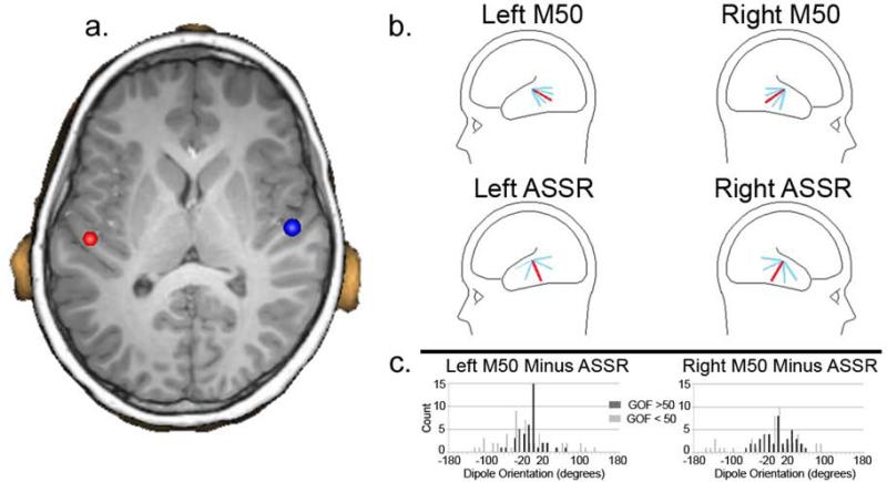
1a) Coronal slice showing placement of dipole sources in left and right Heschl's Gyrus. As detailed in the ‘Results’, a main effect of Hemisphere showed more posterior dipole placements on the left than right (4.09 mm y-axis difference). 1b) Top row: M50 dipole orientation for the left and right hemisphere (total sample) with the average M50 dipole orientation in red and with 1 and 2 standard deviation bars in blue. Bottom row: 40 Hz ASSR dipole. For both the M50 and 40 Hz ASSR, the sagittal dipole angle was examined as this best captures the variability in the tangentially oriented STG M50 and 40 Hz ASSR dipole orientations. 1c) For each participant the difference in the M50 and 40 Hz ASSR dipole orientation was computed for the left and right hemisphere. The histogram shows orientation difference values for participants with better GOF (> 60%; dark gray) and worse GOF (<50%; light gray), and indicates greater similarly (tighter clustering) in individuals with better GOF values, with no participant with a better GOF exceeding a 80 degree (left) or 50 degree (right) difference.

