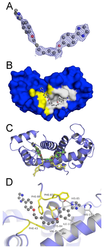
Figure 3. Crystallographic Structure of the S100B•5a Complex (PDB ID: 5DKN).
(A) |Fo|-|Fc| electron density omit map (blue mesh) of 5a (ball and stick) contoured at 2.5σ levels. (B) 5a within Sites 2 (yellow highlight) and 3 (gray highlight) of the S100B homodimer (surface in blue). (C) Modeled orientation of 5a overlaid on the previously elucidated compound-bound structures of pentamidine and heptamidine. (D) The specific interactions of 5a within dimeric S100B (blue ribbons) are rendered with residues (sticks) within 4Å of the 5a highlighted in yellow (Site 2) or gray (Site 3). 5a is situated within a hydrophobic pocket with atoms within hydrogen bond distance at the backbone carbonyl of His85 and the sidechain of Cys84.

