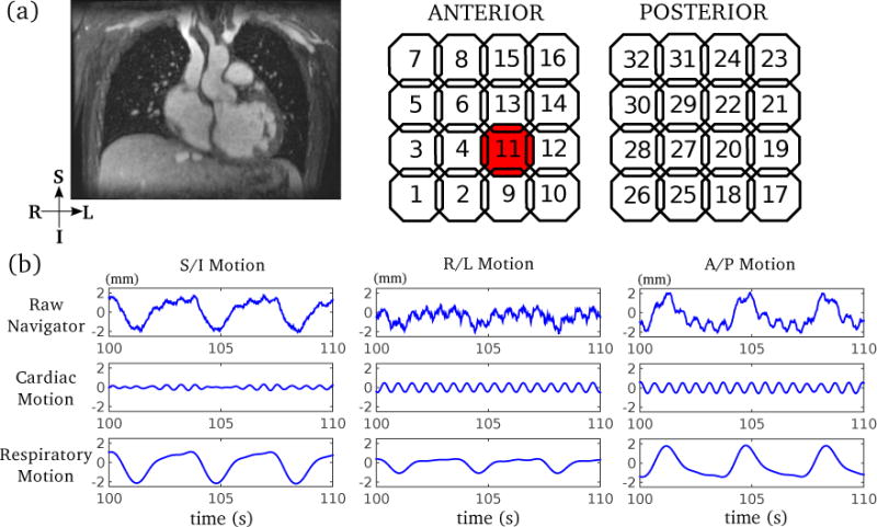
Figure 2.

Example of 3D motion estimates in free-breathing cardiac MRI. (a) Typical imaging volume of a 3D phase-contrast cardiac MRI; (b) 3D motion estimates (navigators) from an example coil element (highlighted in (a)) are shown to demonstrate the separation of cardiac motion and respiratory motion using bandpass filtering. The raw motion estimate contains both cardiac motion and respiratory motion, which can be separated by bandpass filtering prior to coil clustering.
