
Figure 3.
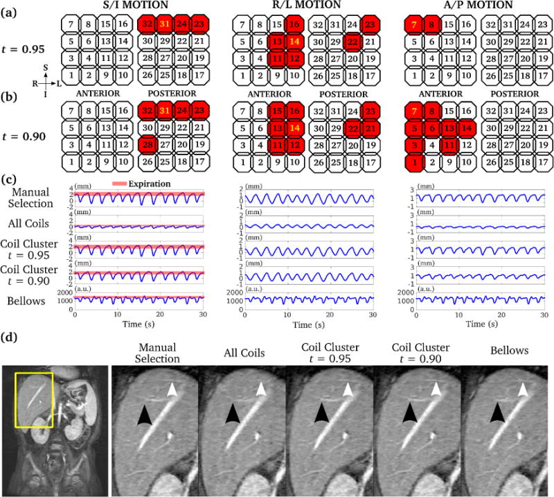
Comparison of 3D motion estimates for abdominal MRI using manual selection, averaging of all coils, coil clustering, and respiratory bellows. Selected coil elements with two different coil clustering parameters (t = 0.95 and 0.90) are highlighted by red in (a) and (b) respectively. The manually selected coil element is highlighted by yellow in (a) and (b). As the threshold t decreased, more coils were selected by coil clustering. The corresponding motion estimates by all methods are shown in (c). Retrospective respiratory gating was performed using each of the motion estimate in (c), and the corresponding images are shown in (d). The proposed coil clustering method achieved very similar image quality compared to that with manual selection, with sharp delineation of branches of the heptic veins (arrows).