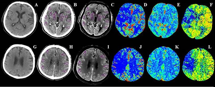
Fig 2. Non-constrast CT, baseline source and average CT perfusion images, cerebral blood flow (CBF), cerebral blood volume (CBV) and mean transit time (MTT) maps at ganglionic (A-F) and supraganglionic (G-L) axial ASPECTS levels.
Multiple circular regions of interest (ROIs) larger than 1 cm2 placed freehand in the affected hemisphere and automatically reflected into homologous regions of the contralateral hemisphere were used to measure CBF, CBV and MTT absolute values from the corresponding functional maps in ASPECTS regions at ganglionic (anterior middle cerebral artery cortex, middle cerebral artery cortex lateral to insular ribbon, posterior middle cerebral artery cortex, insula, caudate nucleus, lentiform nucleus and internal capsule) and supraganglionic (anterior, lateral, and posterior middle cerebral artery cortical territories immediately superior to the previous ones, rostral to basal ganglia) sections.

