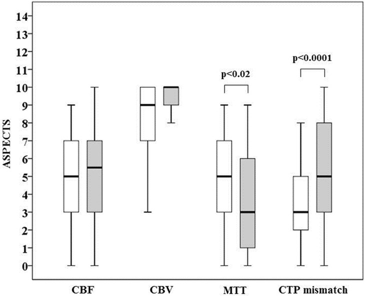
Fig 3. Comparison between semi-quantitative (white boxes) and quantitative (grey boxes) ASPECTS for cerebral blood flow (CBF), cerebral blood volume (CBV), mean transit time (MTT) and CT perfusion (CTP) mismatch.
The boundaries of the box represent the 25th-75th quartile. The line within the box indicates the median. The whiskers above and below the box correspond to the highest and lowest values, excluding outliers.

