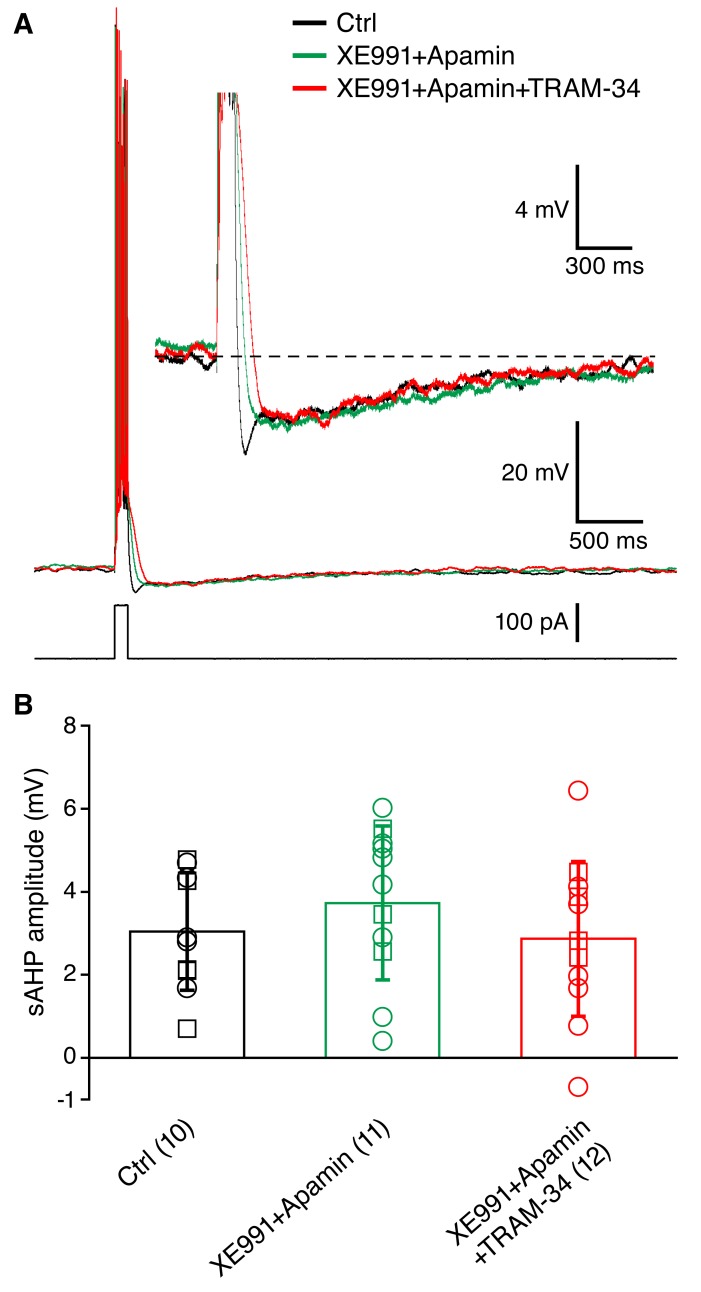
Figure 4. Incubation of acute hippocampal slices and organotypic slice cultures with TRAM-34 (1 μM) did not reduce the sAHP in CA1 pyramidal neurons.
(A) Example traces showing the mAHP and sAHP in three different conditions tested. Cells in the control group (black trace) were recorded in normal ACSF. Cells in the second group (XE991+Apamin, green trace) were recorded after incubation for at least 30 minutes in ACSF with XE991 (10 μM) and apamin (100 nM). Cells in the third group (XE991+Apamin+TRAM-34, red trace) were recorded after incubation for at least 30 minutes in ACSF with XE991 (10 μM), apamin (100 nM) and TRAM-34 (1 μM). (B) Summary of the results from slices (open circles) and organotypic cultures (open squares). The sAHP amplitude did not differ between the three groups of cells: (1) control (n = 10), (2) XE991+Apamin (n = 11), and (3) XE991+Apamin+TRAM-34 (n = 12).

