. 2015 Nov 2;4:e10510. doi: 10.7554/eLife.10510

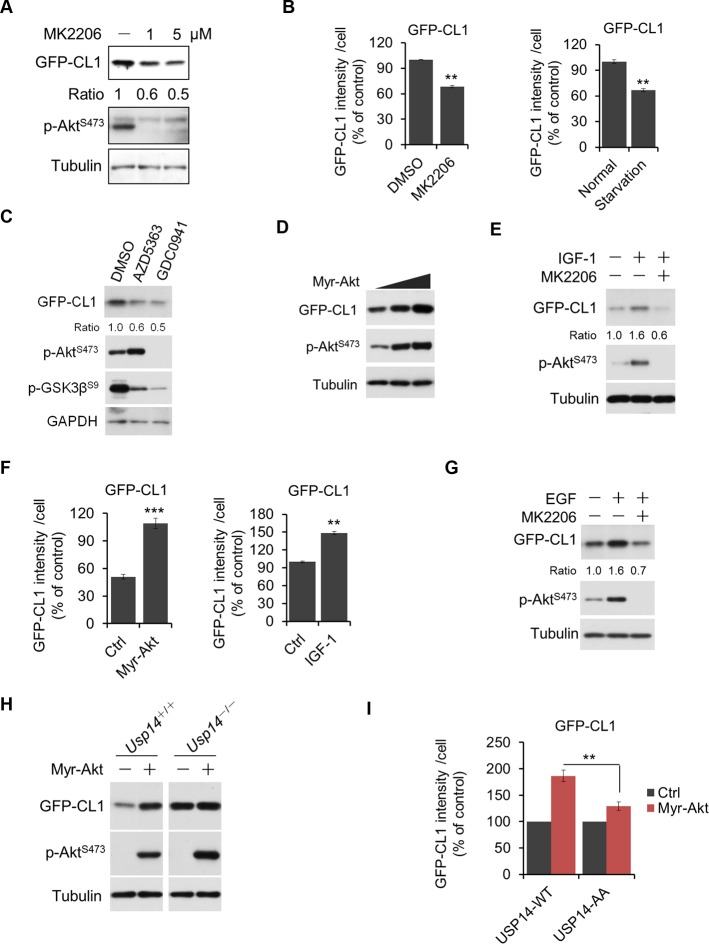
Figure 5. Akt regulates ubiquitin–proteasome system (UPS) function through phosphorylation of ubiquitin-specific protease-14 (USP14).

(A) Inhibition of Akt promotes UPS function. H4-GFP-CL1 cells were treated with different concentrations of MK2206 as indicated for 4 hr. The cells were then harvested and subjected to western blotting analysis using indicated antibodies. (B) H4-GFP-CL1 cells were treated as in (A) and Figure 5—figure supplement 1D. Images of the cells were collected using an ArrayScan HCS 4.0 Reader. The average GFP intensity in 2,000 cells from each indicated sample was determined. Data are displayed as mean ± SD of the GFP intensity per cell. **p<0.01, ***p<0.001 (C) Inhibition of Akt or phosphoinositide 3-kinase (PI3K) promotes UPS function. H4-GFP-CL1 cells were treated with Akt inhibitor AZD5363 (1 μM) or PI3K inhibitor GDC0941 (1 μM) as indicated for 4 hr. The cells were then harvested and subjected to western blotting analysis using indicated antibodies. p-GSK3β(S9) was blotted to indicate the inhibition of Akt. (D) Activation of Akt inhibits UPS. H4-GFP-CL1 cells were transfected with Myr-Akt for 24 hr. The cells were then harvested and subjected to western blotting analysis using indicated antibodies. (E) IGF-1 stimulation inhibits UPS function. H4-GFP-CL1 cells were serum-starved and pretreated with Akt inhibitor MK2206 (1 μM) for 30 min before stimulating with IGF-1 (100 ng/mL) for 30 min. The cells were then imaged and quantified as in (B). (F) H4-GFP-CL1 cells were treated as in (D) and Figure 5—figure supplement 1E. Then cells were imaged and quantified as in (B). (G) EGF stimulation inhibits UPS function. H4-GFP-CL1 cells were serum-starved and pretreated with Akt inhibitor MK2206 (1 μM) for 30 min before stimulation with EGF (100 ng/mL) for 1 h. The cells were then harvested and subjected to western blotting analysis using indicated antibodies. (H) Akt regulates UPS function through USP14. Myr-Akt was transfected into either wild type or Usp14–/– H4 cells stably expressing GFP-CL1 for 24 hr. The cells were then harvested and subjected to western blotting analysis using indicated antibodies. (I) Akt regulates UPS function through phosphorylation of USP14. Myr-Akt was transfected into either wild type USP14 or USP14 AA reconstitution cell lines stably expressing GFP-CL1 for 24 hr. The cells were then imaged and quantified as in (B).

DOI: http://dx.doi.org/10.7554/eLife.10510.012

Figure 5.

Figure 5—figure supplement 1. Regulation of UPS by Akt.

Figure 5—figure supplement 1.

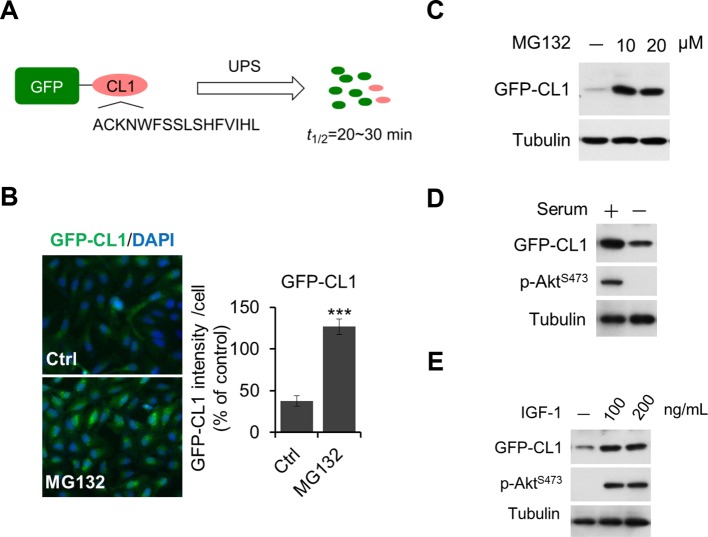
(A, B, C) Validation of GFP-CL1 assay. A schematic representation of GFP-CL1 assay. CL1 degron (ACKNWFSSLSHFVIHL) was added to the C-terminal of GFP as a UPS activity reporter (A). H4-GFP-CL1 cells were treated with 10 μM MG132 for 4 hr. Images of the cells were collected using an ArrayScan HCS 4.0 Reader. The average GFP intensity in 2,000 cells from each indicated sample was determined. The data are displayed as means ± SD of the GFP intensity per cell (B). H4-GFP-CL1 cells were treated with different concentration of MG132 as indicated for 4 hr, and then cells were harvested and subjected to western blotting analysis using indicated antibodies (C). (D) H4-GFP-CL1 cells were serum-starved overnight and harvested and subjected to western blotting analysis using indicated antibodies. (E) H4-GFP-CL1 cells were serum-starved overnight before stimulation with IGF-1 for 30 min. Then the cells were harvested and subjected to western blotting analysis using indicated antibodies.
Figure 5—figure supplement 2. Regulation of UPS by Akt depends on phosphorylation of USP14.

Figure 5—figure supplement 2.

(A, B, C, D) Generation of USP14 knockout H4 cell line using CRISPR/Cas9 system. A schematic of the Cas9/sgRNA/oligo targeting site in the exon2 of Usp14. The sgRNA coding sequence is underlined and labeled in red. The protospacer-adjacent motif (PAM) sequence is underlined (A). The deleted sequences in the Usp14–/– cell lines are presented. The number of sequences analyzed is indicated right (B). Sequencing analysis of Usp14–/– cell lines. The arrow indicates the missing sequences (TCAAG) (C). Western blotting analysis of USP14 expression in WT cells and Usp14–/– cells (D). (E) Akt regulates UPS function through phosphorylation of USP14. An expression vector for Myr-Akt was transfected into either wild type USP14 or USP14 AA reconstitution cell lines stably expressing GFP-CL1 and incubated for 24 hr. Then the cell lysates were harvested and subjected to western blotting analysis using indicated antibodies. (F) GFP-cODC assay was verified. H4-GFP-cODC cells were treated with 10 μM MG132 for 4 hr, and then the cell lysates were harvested and subjected to western blotting analysis using indicated antibodies. (G) H4-GFP-cODC cells were treated with 10 μM MK2206 for 4 hr or transfected with an expression vector for Myr-Akt as indicated for 24 hr. Then the cell lysates were harvested and subjected to western blotting analysis using indicated antibodies.