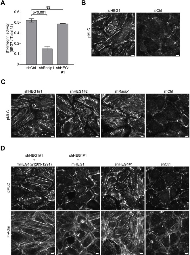
Figure 6. Deletion of the Rasip1-binding site in HEG1 prevents suppression of MLC phosphorylation.
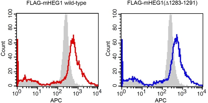
(A) Levels of activated β1 integrin (9EG7) in Rasip1- or HEG1-depleted Human Umbilical Vein Endothelial Cells (HUVEC) were measured by flow cytometry. Depletion of Rasip1 (shRasip1) decreased levels of activated β1 integrin compared to control cells (shCtrl). In contrast, depletion of HEG1 (shHEG1#1) had no effect on levels of activated β1 integrin. Levels of 9EG7 binding were corrected for total β1 integrin expression. Mean values ± SEM of three independent experiments are shown. One-way analysis of variance (ANOVA) with Bonferroni’s test was used to compare each condition with control cells (shCtrl). (B and C) Myosin light chain phosphorylation (pMLC) was analyzed by Spinning Disk Confocal Microscopy (SDCM) in HUVEC, transfected or infected as indicated. siRNA- or shRNA-mediated depletion of HEG1 (B&C; siHEG1, shHEG1#1, shHEG1#2) increased MLC phosphorylation and stress fiber formation. Similarly, lentiviral depletion of Rasip1 (shRasip1) also increased MLC phosphorylation and stress fiber formation. Representative images of 3 independent experiments are shown. Scale bars, 10 µm. (D) Myosin light chain phosphorylation (pMLC) and F-Actin were analyzed by Spinning Disk Confocal Microscopy (SDCM) in Human Umbilical Vein Endothelial Cells (HUVEC), infected as indicated. Lentiviral depletion of HEG1 (shHEG1) increased levels of pMLC and actin stress fiber formation in HUVEC compared to control cells (shCtrl) which was rescued by expression of FLAG-tagged shHEG1#1-resistant wild-type murine HEG1. In contrast, Rasip1-binding deficient murine HEG1(∆1283-1291) (corresponding to aa 1327-1335 in human HEG1) failed to rescue pMLC expression and the increase in actin stress fibers. Expression of rescue constructs, analyzed by flow cytometry, is shown in Figure 6—figure supplement 1. Representative images of 3 independent experiments are shown. Scale bars, 10 µm.


