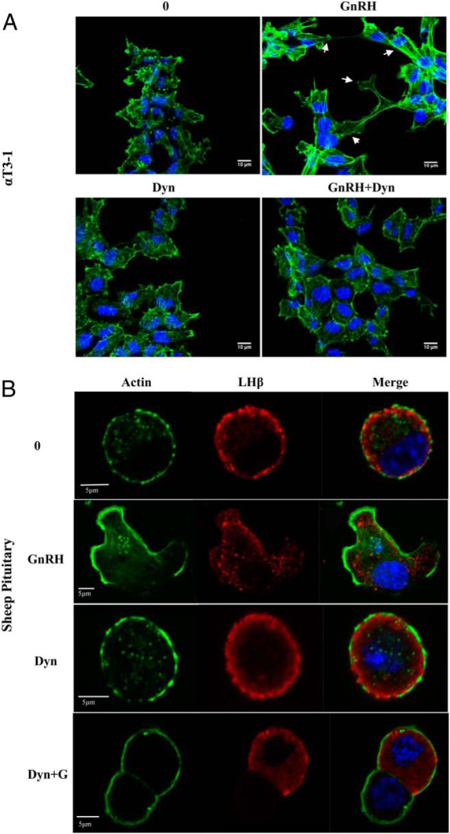
Figure 6.

Dynamin inhibition blunts GnRHa-induced actin remodeling in αT3–1. A, αT3–1 were grown on glass-bottom microwell dishes for 24 hours. Cells were incubated in the presence and/or absence of 80μM dynasore for 30 minutes and/or 10nM GnRHa for 10 minutes followed by fixation in 4% PFA. Cells were then stained with Alexa Fluor 488-conjugated phalloidin (green) and DAPI and imaged by CLSM. The white arrows are highlighting GnRH-induced actin reorganization. B, Dissociated sheep ovine pituitary cells were plated and treated as described in A. After fixation, pituitary cells were immunostained for LHβ followed by the appropriate Alexa Fluor 594-conjugated secondary antibody. Cells were then stained with Alexa Fluor 488-conjugated phalloidin and DAPI and imaged by CLSM.
